|
 1.
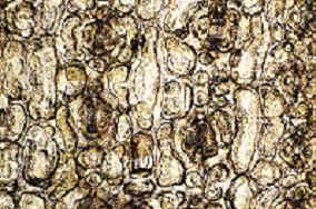
Leaf epidermal cells angular in T-sect. (Fig. 15), papillose on the
abaxial (ventral) 1.
Leaf epidermal cells angular in T-sect. (Fig. 15), papillose on the
abaxial (ventral)
midrib; North America (SE Alaska and W Florida) to Central America
(Honduras,
El Salvador), Himalayas, SW China...............................................
.........................................................................—I. Wallichiana Group (Wallichiana Subgroup)
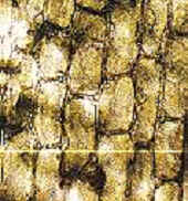
1. Leaf epidermal cells ±elliptical or wide
rectangular in T-sect., 1.5–3.5 times as wide 1.5–3.5 times as wide
as tall (Fig. 16), variable in development of
midrib papillae—2
2. Stomata
bands in dried leaves not sharply differentiated
from adjacent marginal and midrib regions, the abaxial surface
uniformly green to yellowish green or reddish or paler green on
midrib and marginal
zones......................................................................................
—3
2. Stomata
bands distinct in dried leaves by glossy margins and
midrib, the margins and midrib often discolored—reddish (Fig.
17).......................—4
3.
Leaves with resinous cells primarily epidermal; papillae ± equally
developed
across stomata bands and midrib (Fig. 18), often more prominent along
cell walls;
stomata sometimes on midrib, bands with (11-) 13–19 (-21) rows;
E Himalayas to S China
......................................—II.
Wallichiana Group (Chinensis Subgroup) |

 1.5–3.5 times as wide
 reddish or paler green on


